Ca2+ : An Ion of Biological Cybernetics
Created | Updated Oct 4, 2004
by
Felix Bast(formarly, Vadakke Madam Sreejith)
IntroductionThe word 'Cybernetics' was first suggested by father of control engineering, Norbert Weiner in 1947 as the science of control and communication. Applications of this stream have been influencing the control and communication aspects of Biological methods as evidenced by birth of specialized journals like 'Cybernetica', 'Kybernetica', 'Journal of Biological Cybernetics' etc. The word 'Bionics' has been used by a number of scientists to refer to the field of the simulation of biological systems and this too should be thought of, as dealing with important bridge between Biology and Cybernetics.
Ca2+ effectively performs control of various life processes and communicates in molecular level between living cells. The importance of calcium in cell biology was recognized as early as 1882 when Sidney Ringer demonstrated that minute amounts of the divalent ion were necessary to maintain heart muscle contractility [See box 1]. About a century later, physiologists first described calcium as a second messenger in analogy with cyclic adenosine 3',5'-monophosphate (cAMP) which was shown by Sutherland and others during the 1960's to be an important mediator of hormonal responses. The term second messenger implies that calcium is released intracellularly as an intermediate and transient signal produced upon excitation of the cell by external stimuli.
Calcium ions play a major role in controlling the functioning of all cells of the body by acting as carriers of intracellular messages. Cells receive their instructions from the body through hormones and neurotransmitters that bind to receptors on their surface. Many of these messages are relayed by the release of calcium ions from internal stores into the cell cytoplasm, or by opening channels in the membrane of the cell, allowing external calcium to enter the cytoplasm. By these means calcium controls a wide range of cellular functions such as muscle contraction, neurotransmitter and hormone release, metabolism, cell division and differentiation.
Calcium ions are quite remarkable in being able to control numerous processes within the same cell simultaneously [Figure 1]. This ability of calcium to provide a number of signals encoded by the same ion is a result of the way in which calcium levels can be altered in the cell. Outside the cell, free calcium ion concentration is around 1mM, whereas the free calcium ion concentration in the cytoplasm of cells is maintained at around 0.1mM, mainly by ATP powered pumps that pump calcium either out of the cell or into an internal store called the endoplasmic reticulum (E.R.). Because the cytoplasm contains high concentrations of proteins that chelate calcium ions, calcium is able to diffuse only short distances within the cell. This means that a tight control is maintained over the movement of calcium ions in the cytoplasm.
Ca2+ Signals: A Lingua Franca of 'Bionics' at Molecular and Cellular Level.
The control configuration of Ca2+ signaling is negative unity feed back. We can call it as dynamical system, as it possesses memory. Let us explain this concept.
Y(t1) is dependent on the output applied before t= t1, if y(t)1 is its output at t= t1. It can be modeled by an algebraic equation. The output y(t1) at t= t1 depends only on the input ‘u’ applied until that instant, but not later. Hence time varying calcium-signaling system is really a ‘casual system’ and is non-anticipatory.
Ca2+ signaling possesses almost universal biophysical mechanisms. In common with cAMP, the key to Ca2+ activity as a secondary messenger is a rise in the intracellular cytosolic concentration of the ion as a result of hormone/neurotransmitter binding to its receptor at cell membrane [see Figure 1.]. They may enter through Voltage Operated Ca2+ Channels (VOCs) in neurons, Receptor Operated Ca2+ Channels (ROCs) in response to neurotransmitters, or Store Operated Ca2+ Channels (SOCs) which open when the internal stores are emptied of Ca2+. Ca2+ is released by two mechanisms, viz., by the activity of Ins 1,4,5 P3 (Inositol 1,4,5,triphosphate; IP3) on E.R. membrane or by Ryanodine receptors on E.R. membrane. Ins 1,4,5 P3 is produced from Ptd.Ins 4,5 P2 (Phosphatidyl Inositol 4,5 bisphosphate; PIP2) by the enzyme PLC (Phospholipase C). PLC is activated by Gs protein, which in turn is activated by binding with first receptor. Rise in intracellular Ca2+ activates Calmodulin dependent Protein Kinase 2, which in turn will phosphorylate cellular proteins triggering a cell response. There is active research going on in the fields of biophysical mechanisms of Ins 1,4,5 P3 dependent Ca2+ regulatory proteins and Ryanodine receptors on the organelle membrane leading to the release of Ca2+ from intracellular stores to the cytosol.
Some of the Ca2+ E.R. is rapidly taken up by the mitochondria and is then returned to E.R., although most of the stored Ca2+ resides in the lumen of E.R.. But if the mitochondria are overloaded with Ca2+, the result is the abnormal mitochondrial metabolism, which may activate programmed cell death, to be discussed in a later section.
The Space-Time-Modulation Trilogy

The Ca2+ control mechanism depends on 1) the space where Ca2+ concentration is higher or lower, 2) the time through which Ca2+ transients operate and finally 3) the modulation of Ca2+ signals.
Space
Ca2+ can be derived from extracellular as well as intracellular stores. Ca2+ from intracellular stores can be released through channels in E.R. or sarcoplasmic reticulam. When a Ca2+ channel opens, a highly concentrated mass of Ca2+ forms around its mouth and when the channel is closed, it is rapidly dissipated by diffusion. This Ca2+ spark is responsible for signaling repertoire. These signals may be either localized in the vicinity of channels or may generate a global Ca2+ wave, which is intracellular or intercellular. If individual Ca2+ channels of neighboring cells are connected, an intercellular global Ca2+ wave is produced which is responsible for smooth muscle induced Nitric Oxide synthesis in endothelium, glial cell function, wound healing etc. Intracellular Ca2+ wave is responsible for fertilization, cardiac muscle contraction, gene transcription, cell proliferation etc. Membrane excitability, synaptic plasticity etc. are under the control of localized Ca2+ wave.
Time
Usually as an adaptive measure, cells avoid very high concentration of Ca2+ by using low amplitude signals or delivering signals as brief transients. When information has to be relayed over longer time periods, cells use Ca2+ oscillations. The periods of localized and global Ca2+ oscillations differ widely. For example, period of localized Ca2+ oscillation in arterial smooth muscle is 0.1-0.5 seconds; it is 10-60 s for global waves in liver cells, 1-35 minutes for Ca2+ waves in human eggs after fertilization and 10-20 hrs for Ca2+ transients that control cell division.
Modulation
To vary the intensity and nature of physiological output, cells use frequency modulation (FM). Smooth muscle lining of artery can be relaxed by increasing frequency of Ca2+ signals. Calmodulin dependent protein kinase 2 is an important enzyme which catalyses the phosphorylation of cellular enzymes by counting Ca2+ transients, where by depends on it's frequency [See Figure 2].
The key question concerning the repetitive transients is their significance, because the "shape" of the Ca2+ signal is an important factor in the modulation of targets. Oscillations are an elegant and efficient way to transmit Ca2+ signals, while at the same time avoiding deleterious sustained elevations of Ca2+. Some Ca2+ -sensitive events, e.g., muscle contraction or synaptic transmission, require the rapid delivery of a spatially confined signal of high intensity. Other processes require that Ca2+ be delivered in a form that is not strictly localized and is persistent over a longer period. These processes demand that the signal change from elementary to global to permit the prolonged exposure of targets to it.
Amplitude Modulated (AM) Ca2+ signals are generally less reliable than FM. But there is growing evidence that varying the amplitude of Ca2+ signals can activate different genes.
In Fertilization and Development

Intracellular Ca2+ concentration of 50nM with in spermatozoa is an extremely important factor required for fertilization. Using flow cytometry it is found out that in response to agonist progesterone, the Ca2+ concentration in side a sperm, is increased to 195nM, before declining approximately 1/2 of the maximum level with in 2 minutes.
During the interaction of sperm and ovum, a Ca2+ oscillation is produced, which might persist for several hours. It is this oscillation, which triggers stimulation of enzymatic machinery leading to ordered mitosis of zygote. One celled Zygote divides into two when a spontaneous Ca2+ oscillation activates it. This orderly program of mitosis is under the control of two distinct oscillators, viz., Ca2+ levels and oscillating levels of cell division proteins. But the main mechanism is thought to be Ca2+ signals, as it persists when dissociated from cell cycle oscillator. During the development of fertilized ovum, the process by which the concentration of intracellular diffusible Ins 1,4,5 P3 concentration sharply increases with each cell division leading to periodic Ca2+ oscillation is presently unknown. But the study shows that hormones and growth factors produced during cytokinesis activate the enzyme PLC, which catalyses the production of Ins 1,4,5 P3 and Di Acyl Glycerol (DAG) from Ptd Ins 4,5 P2 as described previously.
During the cell differentiation of embryo, Ca2+ signaling contributes to body polarity formation. This is clearly proved by an experiment conducted in zebra fish embryo. When anti Ins 1,4,5 P3 antibodies were incorporated, dorso-ventral specification is altered in progeny.
Ca2+ signaling mechanism is extensively utilized during the differentiation of specific cell types to form germ layers on the gastrulation stage of embryo development. During the formation of muscle, spontaneous Ca2+ transients control assembly of the contractile fibers. Ca2+ is also involved in the development of heart and nervous system. Ca2+ dependent "Nuclear Factor of Activated T-cells" (NF-AT) helps to form cardiac septum (Atrio-ventricular) and semilunar heart valves, during the development of heart. Role of Ca2+ transients with varying frequencies on differentiation of developing amphibian neurons was already established. Migration of neuronal cells is another phenomenon, which is under the control of Ca2+ signals. Primary stages of neuronal reticulation leading to complex neuronal circuits of developing brain are also controlled by Ca2+ control mechanism.
In Neurons
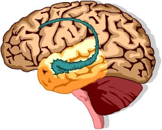
Ca2+ plays a crucial role in neuronal function as well as in information processing of brain. The prime control system of our body itself is under the control of Ca2+. Let us discuss how this happens.
Primarily Ca2+ plays 4 important functions in neurons, viz., signal reception, signal transmission, synaptic plasticity and long term memory.
Neurons have an input zone known as the "dendritic tree", containing several spines, which are responsible for receiving signals from pre-synaptic neuron. When a signal is received (i.e., localized de/pre-polarization due to Na+/K+ ion exchange devise) Ca2+ will enter the spines either from outside (Through VOCs or ROCs) or from internal stores like E.R. (through IP3 operated channels). Hence IP3 and Ca2+ are involved in signal reception. If both IP3 and Ca2+ come together from separate neural signals, it can integrate the information, which is thought to be responsible for learning and memory.
There is growing evidence that local Ca2+ transients are involved in release of neurotransmitter vesicles. Ca2+ can also modulate neuronal sensibility, since it can activate K+ channels leading the efflux of K+ and depressing the nervous transmission.
Neuronal spines are referred to as microprocessors with in the brain, even though it has only 0.1 μm3 volume. It is generally believed that Ca2+ signals with in the spines are responsible for "neuronal plasticity", a phenomenon leading to short-term memory.
Ca2+ is also involved in the consolidation of short-term memory by the genes of nucleus. For triggering the gene transcription far away from the spines, Ca2+ produce other signaling components that migrate in to the nucleus. Thus a long-term memory is created in the genomic level. This is how Ca2+ controls permanent memories.
In Vision Perception
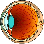
When the rhodopsin is exposed to light, it dissociates to form all-trans retinal and opsin- the visual protein. This conversion also triggers a conformational change to calcium ion channel in the membrane of rod cell. The rapid inflow of Ca2+ triggers a nerve impulse, allowing light to be perceived by the brain.
In Lymphocyte Proliferation

When a specific antigen binds to its receptor site (epitope) on the surface of T or B Lymphocytes, Ins 1,4,5 P3 is produced by unknown mechanisms, which in turn activate the release of Ca2+ from internal stores. In order to compensate decrease in the concentration of Ca2+, these internal stores absorb Ca2+ from ECF through SOCs. This influx of Ca2+ though SOCs is often in the form of regular Ca2+ oscillations that activates factors such as NF-AT, which enter the nucleus causing interactions with operon leading to switch-on of genes. Recently mechanism of immuno-suppression by drugs, such as cyclosporin is discovered to be, by the inhibition of Ca2+ dependent activation of NF-AT. This discovery highlights the relevance of Ca2+ signaling in lymphocyte proliferation.
In Necrosis and Apoptosis
There are several proteases located with in the cell and some of them are sensitive to Ca2+. It is widely accepted that Ca2+ sensitive proteases are directly involved in random cell death - Necrosis.
Ca2+ control mechanism is more relevant in the case of programmed cell death (PCD or apoptosis). The phenomenon of PCD is involved during normal development of tissue patterns as well as in pathological conditions such as AIDS, Cancer and Alzheimer's disease.
Normally most of the stored Ca2+ resides with in the E.R. lumen while the mitochondrion carries very little. This high concentration of Ca2+ inside of E.R. acts not only as store but they are essential for the synthesis and processing of proteins. Stress signals activates shuttling of Ca2+ from E.R. to mitochondria, which in turn leads to 'switch on' of the genes associated with PCD.
Most of the malignant cells contain a mutated protein- Bcl2, which is solely responsible for it's anti-apoptotic activities. Bcl2 prevents cell death, leading to proliferation of cancer cells by modifying the way in which E.R. and mitochondria handle Ca2+ i.e. Bcl2 inhibits the shuttling of Ca2+ from E.R. to mitochondria and thereby prevents Ca2+ depletion of E.R. or Ca2+ excess of mitochondria.
Conclusion
There are many other disciplines of life where Ca2+ mediated control/signal mechanisms particularly appeals. Ca2+ signals control every attributes of life, perhaps with very few exceptions. These ubiquitous and unique properties make Ca2+, an ion of Biological Cybernetics. Regulation of life processes with control of Ca2+ signaling is expected to become as important as genetic engineering with in next few years [Look box 2]. There are multitudes of functions controlled by Ca2+, ranging from initiation of cell division or cellular apoptosis requiring several hours to the sub-second secretory and contractile events. It can be concluded here that the cybernetics of Ca2+ signaling exhibits significant variations in pattern and mechanism of recognition.
Acknowledgements

I thank Council of Scientific and Industrial Research, Govt of India for the support via Junior Research Fellowship Scheme.
Suggested Reading
[1] Clapham, 'Calcium Signaling', Cell, Vol 80, pp.259-268, 1995.
[2] Ramakalyan,A and J R Vengateswaran, 'Systems and Control Engineering - Basic Concepts of Systems', Resonance, Vol 4, No 3, pp.45-52, 1999.
[3] ‘Signal Transduction Knowledge Environment’- an animated web tool to locate signal transduction key pathways, accessible at Science website
[4] Nand K Shah, Tarvinder K Taneja and Seyed E Hasnain, 'Mitochondria can Power Cells to Life and Death - Role of Mitochondria in Apoptosis', Resonance, Vol 5, No. 4, pp.74-84, 2000,
[5] Berridge, M.J, 'Differential activation of transcription factors induced by Ca2+ response, amplification and duration', Nature, Vol 386 No 6627, pp.855-858,1997.
[6] Berridge, M. J., 'Neuronal calcium signaling', Neuron Vol 21, pp.13-26, 1998.
[7] D. H. Mac Lennan and N. M. Green, 'Pumping ions', Nature, Vol 405, p.633, 2000.
[8] J.W. Putney, Jr., 'Calcium Signaling: Up, Down, Up, Down…. What's the Point?', Science, Vol 279, pp.191 - 192, 1998.
[9] 'Molecular Cell Biology' (4th edn.), Harvey Lodish and others, W.H.Freeman and Co., Part 4, Chapter 20, 2002.
Box1. "The London Tape Water Mystery"

It was 120 years ago, when Sidney Ringer was studying the contraction of isolated rat hearts… an experiment which by today's standards would be considered unacceptably sloppy, marked the beginning of the Ca2+ signaling saga [Ringer, S., J. Physiol. Vol. 4, pp. 29-43, 1883]. In earlier experiments, Ringer had suspended them in a saline medium for which he admitted to having used London tap water, which is hard: The hearts contracted beautifully. When he proceeded to replace the tap water with distilled water, he made a startling finding. The beating of the hearts became progressively weaker, and stopped altogether after about 20 min. To maintain contraction, he found it necessary to add Ca2+ salts to the suspension medium. Thus, Ringer had serendipitously discovered that Ca2+ hitherto exclusively considered as a structural element, was active in a tissue that has nothing to do with bone or teeth, and performed there a completely novel function: It carried the signal that initiated heart contraction. It was a landmark observation, which should have immediately aroused wide interest. Unexpectedly, however, for decades it attracted no particular attention. Occasionally, farsighted pioneers argued forcefully for a messenger role of Ca2+, offering compelling experimental evidence. Among them, one could quote L. V. Heilbrunn [Heilbrunn, L. V., Physiol. Zool. Vol. 13, pp. 88-94, 1940], who contracted frog muscle fibers by applying Ca2+ salts to their cut ends, but not to their surfaces. Heilbrunn correctly concluded that Ca2+ had diffused from the cut ends to the internal contractile elements to elicit their contraction. One could also quote K. Bailey [Bailey, K. Biochem. J. Vol. 36, pp. 121-139, 1942], who showed that the ATPase activity of myosin was strongly activated by Ca2+ (but not by Mg2+), and concluded that the liberation of Ca2+ in the neighborhood of the myosin controlled muscle contraction. Clearly, enough evidence was there, but only a handful of people had the vision to see it and to foresee its far-reaching implications. Perhaps O. Loewy can offer no better example of clairvoyance than the quip in 1959: "Ja Kalzium, das ist alles!"
Box 2. Drugs regulating Ca2+ signaling.
Generally, calcium related chemotherapeutic agents that are in widespread use today target outer membrane channels, which allow calcium to enter the cells from outside. Drugs like verampil block influx of Ca2+ through channels, and hence can be effectively administered for the relaxation of muscles lining blood vessels in patients with hypertension, or in angina patients as it enhances blood flow in coronary arteries. As these drugs reduces level of Ca2+ in cardiac muscle tissues, heart became less excitable with normal action potentials and hence patients with dysrhythmias (the irregular heart beating which may lead to heart attack) can be treated with this kind of drugs.
Other group of drugs block calcium pumps or Ca2+ channels - both situated inside the cells - which release Ca2+ from the internal stores. The pump blocker thapsigargin and channel blockers such as xestospongin have proved to be invaluable tools in unlocking the secrets of calcium signaling pathways. However, agents of this type have not been used therapeutically (although thapsigargin is an ancient folk remedy for arthritis). The reason for the lack of pump and internal channel blockers in the therapeutic arsenal may be related to another role played by calcium in the life of cells.
